
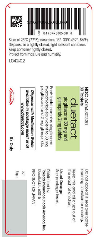
Duetact dosages: 17 mg, 16 mg
Duetact packs: 30 pills, 60 pills, 90 pills, 120 pills, 180 pills, 270 pills, 360 pills
Purchase duetact with mastercard
Scientists have since discovered that many alternative tissues in the physique respond to interleukins. Its main function is to mediate the inflammatory response, however it also has widespread systemic results on immune function and metabolism. Altering blood vessel endothelium to ease passage of white blood cells and proteins during the inflammatory response. The complement cascade terminates with the formation of membrane attack advanced, a bunch of lipid-soluble proteins that insert themselves into the cell membranes of pathogens and virus-infected cells and form big pores (fig. All lymphocytes secrete cytokines that act on immune cells, on non-immune cells, and, typically, on pathogens. Cytokines released by the inflammatory response attract lymphocytes to the site of an immune reaction. Bradykinin Kinins are a bunch of inactive plasma proteins that take part in a cascade similar to the coagulation cascade [p. The finish product of the kinin cascade is bradykinin, a molecule that has the identical vasodilator effects as histamine. Bradykinin also stimulates ache receptors, creating the tenderness related to inflammation. The complement proteins are secreted in inactive types that are activated because the cascade proceeds. Intermediates of the complement cascade act as opsonins, chemical attractants for leukocytes, and agents that trigger mast cell degranulation. Active immunity happens when the body is uncovered to a pathogen and produces its personal antibodies. Active immunity can occur naturally, when a pathogen invades the physique, or artificially, as when we are given vaccinations containing useless or disabled pathogens. The transfer of antibodies from mom to fetus throughout the placenta is one instance. Travelers going abroad may be injected with gamma globulin (antibodies extracted from donated human plasma), however this passive immunity lasts solely about three months because the injected proteins degrade and are cleared from the circulation. Lymphocytes Mediate the Acquired Immune Response On a microscopic level, all lymphocytes look alike. At the molecular level, however, the cell varieties could be distinguished from one another by their membrane receptors. All lymphocytes that bind that ligand kind a gaggle known as a clone klon, a twig (fig. If each pathogen that entered the physique needs a dedicated sort of lymphocyte, there must be millions of various sorts of lymphocytes able to fight hundreds of thousands of various pathogens. But how can the body retailer both the number and number of lymphocytes wanted for sufficient defense As it seems, the immune system keeps only some of every type of lymphocyte available. If the pathogen these cells struggle seems, the cells reproduce to present the numbers wanted. The newly shaped lymphocytes in an expanded clone differentiate into effector cells and memory cells. Second and subsequent exposures to the antigen activate the memory cells and cause rapid clonal enlargement, making a faster and stronger secondary response to the antigen. Antibodies are also known as immunoglobulins, and this alternative name describes what the molecules are: globular proteins that participate within the humoral immune response. When a clone of B lymphocytes responds to antigen exposure, a variety of the effector cells differentiate into plasma cells. Instead, they synthesize and secrete extra antibody molecules at incredible rates, estimated to be as excessive as 2000 molecules per second! Plasma cell antibodies kind humoral immunity, the soluble antibodies of the plasma. After each invader has been efficiently repulsed, the shortlived plasma cells die off-it could be harmful to have them continuing to secrete antibody after the antigen is gone.
16mg duetact otc
Type 4 hypersensitivity, additionally referred to as delayed hypersensitivity, includes antigen�T cell�macrophage interactions figuring out the formation of a granuloma. The Mantoux reaction within the tuberculin pores and skin take a look at is a classic delayed hypersensitivity response. This native reaction is manifested by erythema and edema in the injected skin web site within 48 hours. A persistent granuloma represents an amplified tissue reaction that develops in response to a sustained immune response to released antigens somewhat than to the triggering pathogen itself. Helper T cells or cytotoxic T cells, macrophages and multinucleated giant cells are the hallmark of continual granulomas. We come back to Type four hypersensitivity and continual granuloma once we handle the method of persistent inflammation. Complement offers a fast and environment friendly mechanism for eliminating pathogens to prevent tissue damage or continual infection. Host tissues have cell surface�anchored regulatory proteins, which may inhibit complement activation and prevent unintended injury. The complement system consists of about 20 plasma proteins, synthesized mainly in the liver, that complement, or improve, a tissue response to pathogens. Binding of mannose-binding lectin to a bacterial carbohydrate moiety (lectin pathway). By spontaneous activation of C3, a proenzyme (inactive precursor) of the complement sequence (alternative pathway). The crucial molecule of the complement cascade is C1, a hexamer, referred to as C1q, with binding affinity to the Fc area of an immunoglobulin. When the globular domains of C1q bind to the Fc areas of immunoglobulins already certain to the surface of a pathogen, C1r is activated and converts C1s right into a serine protease. The third step happens when complement protein C2 is cleaved by C1s into C2a (discarded) and C2b. C2b binds to the already certain C4b, forming the complicated C4b-2b, additionally called C3 convertase, on the floor of a pathogen. The fourth step takes place when complement protein C3 is cleaved by C3 convertase into C3a (discarded) and C3b. The C4b-2b-3b advanced, now designated C5 convertase, cleaves complement protein C5 into C5a (discarded) and C5b. The final steps consist within the binding of the opsonized pathogen to complement receptors on the surface of the phagocyte. Complement system C1q C1s C1 is the primary element of the complement activation pathway. Nomenclature the letter "C" followed by a quantity designates the components of the complement cascade. The merchandise of the cleavage of C1, C2, C3, C4, C5, and others are designated by lowercase letters: "a" is the small fragment; "b" is the bigger fragment. C3a and C5a are proinflammatory fragments, which recruit leukocytes to sites of infection and activate them. This conversion generates a serine protease that initiates the complement cascade. It provides a speedy and efficient means for eliminating pathogens and triggering irritation. The complement system has the following specific characteristics essential to bear in mind: 326 10. Complement fragments C3a and C5a, produced by the enzymatic cascade, have proinflammatory exercise. Complement fragments C3a and C5a recruit leukocytes to the infection site, which become activated and activate different cells. General Pathology: Inflammation Invading pathogens (bacteria, virus, parasites and foreign objects) could cause localized tissue damage resulting in an inflammatory response. If the injury persists and the destruction of the tissue (necrosis) continues, an immune response develops with the characteristics of continual inflammation. When a pathogen persists and an an infection course of occurs, tissue destruction continues and concurrent immune responses and fibrous restore take place by the method of chronic irritation. Endothelial cells release nitric oxide to relax the sleek muscle cells of the blood vessel wall to increase blood flow. Enhanced vascular permeability of capillaries and venules ends in the accumulation of fluid, or exudate, in the interstitial space, resulting in tissue swelling.

Order cheapest duetact and duetact
It is equal to atmospheric pressure at the beginning and finish of inspiration and expiration. When lung quantity is at its minimal, alveolar strain is (c) transferring from maximum to minimal and exterior intercostal muscle contraction is (b) minimal. Slow and deep: whole pulmonary air flow = 6000 mL/min, 600 mL contemporary air, alveolar ventilation = 4800 mL/min. This constricts native arterioles, which then shunts blood to better-perfused sections of lung. Nose and mouth, pharynx, larynx, trachea, main bronchus, secondary bronchi, bronchioles, epithelium of the alveoli, interstitial fluid, and capillary endothelium 6. Capillary endothelium is nearly fused to the alveolar epithelium, and the area between alveoli is nearly filled with capillaries. Right ventricle to pulmonary trunk, to left and proper pulmonary arteries, smaller arteries, arterioles, capillaries, venules, small veins, Appendix A Answers A-23 eight. Particles are trapped within the mucus and moved towards the pharynx by mucocilliary escalator. In obstructive lung diseases corresponding to bronchial asthma, the bronchioles collapse on expiration, trapping air within the lungs and leading to hyperinflation. Her larger tidal volume may be the end result of the hassle she should exert to breathe. Increasing both fee and depth has the most important impact and is what would occur in real life. Alveolar ventilation-volume of air entering or leaving alveoli in a given time frame. Bronchoconstrictors: histamine, leukotrienes, acetylcholine (muscarinic); bronchodilators: carbon dioxide, epinephrine (b2) 18. The dead space increases since the person inhales air left behind from the earlier exhalation. Intrapleural pressure has to be more subatmospheric as a outcome of a higher strain gradient is required for expansion. Blood swimming pools within the lungs as a result of the left heart is unable to pump all of the blood coming into it from the lungs. There is a most air flow rate, and the slope of the curve decreases as it approaches this maximum. Pressure gradients, solubility in water, alveolar capillary perfusion, blood pH, temperature. Dorsal-neurons for inspiration; ventral-neurons for inspiration and energetic expiration. Central pattern generator- group of neurons that work together spontaneously to control rhythmic contraction of certain muscle groups. Partial strain and Appendix A Answers A-25 ChApter 19 Concept Check Questions 1. Osmotic strain is higher in efferent arterioles due to similar amount of protein in a smaller volume. Composed of macula densa cells within the distal tubule and granular cells in arteriole wall. Reabsorbed molecules go into the peritubular capillaries and the systemic venous circulation. Excretion is also bulk move however includes motion from the kidney lumen to the skin world. Toilet coaching allows higher brain facilities to inhibit the reflex till an appropriate time. Bladder easy muscle contracts beneath parasympathetic management, so blocking muscarinic receptors decreases bladder contraction. This will permit diffusion of solutes and water from the blood into the dialysis fluid, but diffusion will cease at the desired focus. To take away extra water from the blood, you might make the dialysis fluid more concentrated than plasma. The line might be a straight line starting at the origin and lengthening upward to the right. Location: lie on both aspect of the spine, between the peritoneum and the bones and muscular tissues of the back, and behind the peritoneal cavity 2.
Duetact 16 mg visa
A minor defect of the tight junction barrier can allow bacterial merchandise or dietary antigens to cross the epithelium and enter the lamina propria. Intestinal tight junction barrier Intestinal lumen Claudin Epithelial cell layer 1 Antigens Bacteria Increased flux throughout tight junctions Food Intestinal tight junction barrier 1 A defect within the intestinal tight junction barrier allows the unrestricted passage of antigens to the lamina propria. Therefore, these structures serve important features that may lead to inflammation or tolerance. The lymphoid follicles, every showing a germinal center and a subepithelial dome space. High endothelial venules, enabling the immigration of lymphocytes, are current in the lymphoid follicles. M cells type intraepithelial pockets, the place a subpopulation of intraepithelial B cells resides and express IgA receptors allowing the capture and phagocytosis of IgA-bound micro organism. Dendritic cells migrate to mesenteric native lymph nodes to additionally elicit immune responses. In the lumen, the secretory element is cleaved from its transmembrane anchorage. The population of M cells will increase quickly within the presence of pathogenic bacteria within the intestinal lumen (for instance, Salmonella typhimurium). When confronting Salmonella, the microfolds of M cells change into giant ruffles and, inside 30 to 60 minutes, M cells undergo necrosis and the population of M cells is depleted. The subepithelial dome contains B cells, T cells, macrophages, and dendritic cells. Intestinal antigens, certain to immunoglobulin receptors on the surface of B cells, interact with antigen-presenting cells at the subepithelial dome region. Polymeric IgA Plasma cells secrete polymeric IgA into the intestinal Polymeric IgA 16. Intestinal mucus blanket In the intestine, goblet cells secrete mucin glycoproteins that type a blanket consisting of stratified outer and inner layers on the surface of the epithelium. Microorganisms predominate within the outer mucus layer, whereas the inside mucus layer, immune to microorganism penetration, accommodates antimicrobial proteins secreted by Paneth cells and enterocytes. Enteroendocrine cell Muscularis mucosae Paneth cell Lymphocytes Enteroendocrine cell lumen, the respiratory epithelium, the lactating mammary gland, and salivary glands. Most plasma cells are present within the lamina propria of the intestinal villi, together with lymphocytes, eosinophils, mast cells, and macrophages. Polymeric IgA is secreted as a dimeric molecule joined by a peptide referred to as the J chain. Polymeric IgA binds to a particular receptor, known as the polymeric immunoglobulin receptor (pIgR), available on the basal surfaces of the enterocytes. The polymeric IgA�pIgR�secretory component advanced is internalized and transported throughout the cell to the apical floor of the epithelial cell. At the apical floor, the complex is cleaved enzymatically and the polymeric IgA-secretory element complex is released into the intestinal lumen 514 sixteen. IgA attaches to bacteria and soluble antigens, preventing a direct damaging effect to intestinal cells and penetration into the lamina propria. One final point: IgA regulates the composition and the function of the intestinal microbiota by affecting bacterial gene expression. Lower half of an intestinal gland (crypt of Lieberk�hn) Plasma cell Lumen of a lacteal Mitotically dividing stem cells Brush border Paneth cells Nucleus Muscularis mucosae Enterocytes Paneth cells 16. Glands of Lieberk�hn 1 Photomicrograph from Cotran R, et al: Robbins Pathologic Basis of Disease, sixth ed. Paneth cells Enterocytes and Paneth cells in particular secrete proteins to restrict micro organism pathogenic challenges. We continue the discussion inside the context of the antimicrobial defense of the intestinal mucosa involving Paneth cells and enterocytes. By making a barrier that limits direct entry of luminal bacteria to the epithelium. Paneth cells are current on the base of the crypts of Lieberk�hn and have a lifetime of about 20 days. The pyramid-shaped Paneth cells have a basal domain containing the tough endoplasmic reticulum.
Order 16 mg duetact free shipping
Define and relate each of the following terms in each group: (a) gamete, zygote, germ cell, embryo, fetus (b) coitus, erection, ejaculation, orgasm, emission, erogenous zones (c) capacitation, zona pellucida, acrosomal response, cortical response, cortical granules (d) puberty, menarche, menopause, andropause 20. The babies of moms with gestational diabetes mellitus are likely to weigh extra at start. The following graph exhibits the outcomes of an experiment by which regular men were given testosterone over a period of months (indicated by the bar from A to E). The rate of increase is about the identical in women and men, indicated by the nearly parallel traces. To maintain mass stability, both metabolites have to be either excreted or additional metabolized. To present the distinction between men and women, the graphs ought to have both separate traces or separate bars for men and women. Physiology studies the normal functioning of an organism, while anatomy research the buildings inside the organism. Stimulus, sensor, enter signal, integrating center, output signal, goal, response. Evaluate your map by evaluating it to some done by classmates or to ask your instructor for feedback. You would possibly embody completely different sorts of breads, meats, and so forth, or add a class of sandwich characteristics, corresponding to temperature (hot, cold) or layers (single, club). Exchange with external environment-respiratory exchanges gases; digestive system takes in nutrients; digestive and urinary remove waste products. Negative feedback-feedback signal turns response loop off; helps keep homeostasis. For peer critiques of the examine, see New England Journal of Medicine 347(2): 132�133 and 137�139, 2002, July 11. Molecule B is a greater candidate because its smaller Kd means higher binding affinity. The 5 g of glucose add quantity, so when you begin with one hundred mL of solvent, you end up with more than a hundred mL of answer. Each double bond removes two hydrogens from the molecule, due to this fact, (c) C18H30O2 is essentially the most unsaturated as a outcome of it has the fewest hydrogens. A nucleotide consists of one or more phosphate groups, a 5-carbon sugar, and a nitrogenous base. Soluble proteins act as enzymes (for metabolic reactions), immunoglobulins, signal molecules, binding proteins, regulatory proteins, receptors, and membrane transporters (carriers or ion channels). If the drug is performing as a competitive enzyme inhibitor, then its binding would be reversible; growing the focus of the substrate would displace the competitive inhibitor and decrease the inhibition. Myoglobin has a better affinity for O2 as a outcome of at decrease oxygen concentrations, myoglobin binds more O2 than hemoglobin does. Proteins could additionally be transmembrane, lipid-anchored, or loosely bound to other proteins. To hide the hydrophobic tails of phospholipids from direct contact with aqueous body fluids 4. Cytosol is the semi-gelatinous substance by which organelles and inclusions are suspended. Cilia are brief, often are very numerous on a cell, and transfer fluid or substances throughout the cell surface. Flagella are longer, normally happen singly on human sperm, and are used to propel a cell through a fluid. The membranes of organelles create compartments that physically isolate their lumens from the cytosol. The double membrane of mitochondria creates two different compartments contained in the organelle. It suggests that the cell has a excessive power requirement as a end result of mitochondria are the location of best energy manufacturing in the cell. This suggests that the tissue synthesizes massive amounts of lipids, fatty acids, or steroids, or that it detoxifies international molecules. Collagen provides strength and flexibility; elastin and fibrillin provide elastance; fibronectin helps anchor cells to matrix. Cartilage lacks a blood provide, so oxygen and nutrients needed for restore should attain the cells by diffusion, a gradual process.
Cheap 17mg duetact
The radioactivity is detected by coating the cells with a skinny layer of a photographic emulsion. After improvement of the emulsion, silver grains indicate the location of the labeled constructions. This approach has been used extensively for determining the duration of a number of phases of the cell cycle. Regulation of the cell cycle 4 G2/Mitosis transition: cyclin A/Cdk1 activity is required for the initiation of prophase. The phosphorylation of Rb protein is accomplished resulting in further activation of E2F-mediated transcription. Cdk2 Cyclin A Early G1: Cdk4 and/or Cdk6 are activated by cyclin D and initiate the phosphorylation of retinoblastoma (Rb) protein. This determines the release of E2F transcription components activating cyclin E and cyclin A genes. Cyclin E Cdk2 Cyclin E Cyclin A Phosphorylated Rb protein Release of E2F transcription factors 1. Cell progress is required for doubling the cell mass in preparation for cell division. In a extra contemporary view, the cycle is regarded as the coordinated development and completion of three separate cycles: 1. A cytoplasmic cycle, consisting of the sequential activation of cyclin-dependent protein kinases in the presence of cyclins. A centrosome cycle, consisting of the duplication of the 2 centrioles, referred to as mom and daughter centrioles, and meeting of pericentriolar 44 1. Recall from our previous dialogue on the centrosome as a microtubule organizing middle that -tubulin ring complexes are microtubule-nucleating complexes interacting with the protein pericentrin within the pericentriolar material. If this interplay is disrupted, the cell cycle is arrested through the G2-M part transition, and the cell undergoes programmed cell death or apoptosis. The activities of cyclin-dependent protein kinases� cyclin complexes coordinate the timed development of the nuclear and centrosome cycles. Cells can be stained through the developed emulsion layer to decide the exact localization sites of the overlapping silver grains. The time development of cells through the different phases of the cell cycle can be estimated utilizing both transient and extended [3H]thymidine pulses. The variety of cells radiolabeled during interphase (generally about 30%) symbolize the labeling index of the S part. Assembly and disassembly of the nuclear envelope 1 During interphase, the nuclear lamina, a network of lamins A, B, and C, associates with chromatin and the inside membrane of the nuclear envelope. Inner nuclear membrane Nuclear lamina Chromatin 2 At mitosis, first protein kinase C and then cyclin A�activated Cdk1 kinase phosphorylate lamins, inflicting the filaments to dissociate into free lamin dimers. Head Lamin dimers Rod Tail Phosphorylation web site 3 As the nuclear lamina dissociates, the nuclear envelope undergoes breakdown. Cisternae of the endoplasmic reticulum are a reservoir of the longer term nuclear envelope. Telophase Cisterna of the endoplasmic reticulum related to chromatin Fragmented cisternae of the endoplasmic reticulum Endoplasmic reticulum cisternae Chromosome Phosphorylated lamins A, B, and C Dissociated nuclear pore complicated Sequential occasions during the reassembly of the nuclear envelope four During anaphase, soluble proteins of the nuclear pore advanced (nucleoporins) bind to the floor of chromatin. Before cytokinesis, lamin B turns into dephosphorylated by protein phosphatase 1 and, along with lamins C and A, initiates the formation of the nuclear lamina. The formation of the nuclear lamina begins on completion of the reconstruction of the nuclear envelope. Breakdown and reassembly of the nuclear envelope S G1 Cdk4 Cyclin D Restriction level Phosphorylated Rb protein, by the action of the cyclin D�Cdk4 complex, facilitates the passage via the restriction level. Unphosphorylated Rb protein prevents progression of the cell cycle past the restriction point in G1 mitosis (mitotic index) indicates that the radiolabeled precursor, which entered the cell through the S part, progressed via the G2 part into M phase. The nuclear lamina consists of kind V intermediate filament proteins, lamins A, B, and C, which affiliate with one another to kind the nuclear lamina. Phosphorylation of lamins, catalyzed first by protein kinase C and later by cyclin A�activated Cdk1 kinase, results in the disassembly of the nuclear lamina. In addition, the elements of the nuclear pore advanced, the nucleoporins, and the membranous cisternae of the endoplasmic reticulum additionally disperse. The endoplasmic reticulum is the nuclear membrane reservoir for nuclear envelope reassembly. During anaphase, nucleoporins and three transmembrane protein parts of the inside nuclear membrane, lamina-associated polypeptide 2, lamin B receptor, and emerin, attach to the floor of the chromosomes (chromatin).
Buy cheap duetact 16 mg line
When Ca2+ ranges lower, the myosin gentle chain is enzymatically dephosphorylated, and the muscle relaxes. Concept mapping Muscle tissue Muscle Tissue Skeletal muscle Cardiac muscle Cardiac muscle cell/cardiocyte (central nucleus) Purkinje fibers Myofibril Intercalated disk Actin Smooth muscle Smooth muscle cell (central nucleus) General organization Epimysium Perimysium Endomysium Microscopic group Satellite cell Neuromuscular spindle Skeletal muscle cell (multinucleated, peripheral nuclei) Intrafusal fibers Myofibril Sarcomere Motor finish plate Myofilaments Z disks Actin Nebulin Titin Myosin Neuromuscular junction Intermediate Myosin filaments Sarcomere Transverse and longitudinal elements Z disks Myofilaments Diad (at the Z disk) Triad (at the A-I junction) Essential ideas Muscle Tissue Each skeletal muscle cell is surrounded by a plasma membrane (called sarcolemma). The sarcolemma initiatives long processes, called transverse tubules or T tubules, deep into the cytoplasm (called sarcoplasm). Each T tubule is flanked by sacs of the endoplasmic reticulum (called sarcoplasmic reticulum) forming a tripartite structure referred to as a triad, found on the junction of the A band and I band. The association of these two myofilaments generates a banding sample (or striation), characteristic of skeletal and cardiac muscle tissue. Actin is associated with the tropomyosinEssential ideas � There are three kinds of muscle: (1) Skeletal muscle. Skeletal muscle is surrounded by the epimysium, a layer of dense connective tissue. The perimysium, derived from the epimysium, surrounds bundles or fascicles of muscle cells, additionally referred to as muscle fibers. Each muscle fiber within a fascicle is surrounded by the endomysium, a skinny layer of reticular fibers and extracellular matrix closely associated to a basal lamina enveloping every muscle cell. Skeletal muscle cells are multinucleated cells, ensuing from the fusion of myoblasts. The desmin-plectin advanced varieties a lattice with the alternative ends attached to costameres within the sarcolemma. This association stabilizes the myofibrils within the sarcoplasm throughout muscle contraction. The size of the sarcomere decreases because actin and myosin slide past one another, represented by a reduction in the width of the I band and H band. Inside the muscle, a motor nerve gives rise to quite a few branches, every innervating a single muscle cell. An excitation-contraction signal is produced by the discharge of acetylcholine from a presynaptic button into a primary synaptic cleft, an invagination on the floor of a muscle cell coated with basal lamina containing acetylcholinesterase. The major synaptic cleft varieties secondary synaptic clefts, additionally covered by basal lamina. An action potential depolarizes the sarcolemma, and the action potential travels inside the muscle cell alongside T tubules, that are in contact with channels of the sarcoplasmic reticulum containing calcium. Calcium ions are released, bind to troponin C, and provoke contraction by regulating myosin-actin interplay. When depolarization ends, calcium ions are pumped again into the sarcoplasmic reticulum channels and bind to calsequestrin. Botulinum toxin binds to the presynaptic membrane of the nerve terminal and blocks the discharge of acetylcholine. Curare binds to the acetylcholine receptor, prevents binding of acetylcholine, and induces muscle paralysis. In myasthenia gravis, an autoimmune disease that produces fatigue with exercise, autoantibodies bind to the acetylcholine receptor and forestall binding of acetylcholine. They present mechanical stabilization throughout muscle contraction: (1) Dystroglycan complicated consists of dystroglycan- and dystroglycan-. Dystroglycan- binds to the chain of laminin-2, and dystroglycan- binds to dystrophin. Sarcoglycanopathies (for instance, limb-girdle muscular dystrophies) are caused by defects in parts of the sarcoglycan advanced. Activated satellite cells activated by trauma or mechanical stress can self-renew and proliferate. The expression of myogenic regulatory elements (for example, Myf5 and MyoD) prompts satellite tv for pc cells, which become myogenic precursor cells (to form muscle cells) or side-population cells (to differentiate into hematopoietic cells). It is provided by sensory and motor nerves and consists of specialised muscle fibers. Muscle fibers on the interior of the neuromuscular spindle are known as intrafusal fibers to distinguish them from the nonspecialized extrafusal fibers, regular skeletal muscle fibers aligned in parallel with the intrafusal fibers.
References
- Brown JW. Aphasia, apraxia, and agnosia. Springfield, IL: Charles C Thomas; 1972.
- Aragona F, Ragazzi R, Pozzan GB, et al: Correlation of testicular volume, histology and LHRH test in adolescents with idiopathic varicocele, Eur Urol 26:61n66, 1994.
- Acharya JN, Pacheco VH. Neurologic complications of hepatitis C. Neurologist 2008;14:151-6.
- Bench T, Burkhoff D, OíConnell JB, et al: Heart failure with normal ejection fraction: Consideration of mechanisms other than diastolic dysfunction, Curr Heart Fail Rep 6:57-64, 2009.