Super P-Force dosages: 160 mg
Super P-Force packs: 10 pills, 20 pills, 30 pills, 60 pills, 90 pills, 120 pills, 180 pills
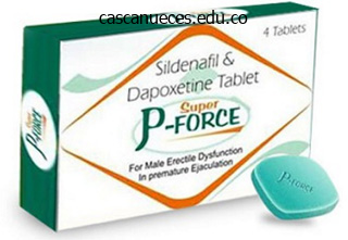
Purchase generic super p-force line
Wrapping the positioning with an elastic bandage for a couple of minutes is suitable, but longer application in this broad method might result in the complete graft clotting. Dialysis Clamps Special self-retaining dialysis clamps could additionally be applied over a selected bleeding site. Apply topical hemostatic brokers instantly over the location of bleeding and hold them in place with gloved hands and a gauze bandage or clamp. If using a skin adhesive, apply the adhesive over the site of bleeding whereas also making use of direct strain proximally. This dialysis graft clamp with sterile gauze (option: impregnate the gauze with topical thrombin) is kept in place for about 10 minutes. Bleeding after dialysis is related to needle size and the degree of anticoagulation, however extended bleeding might sign outflow stenosis, infection, or pores and skin atrophy and the necessity for evaluation of the shunt. Vasoconstrictive Agents Subcutaneous injection of two to four mL of lidocaine (2%) with epinephrine to kind a wheal across the website might decrease bleeding by both vasoconstriction and native stress. Do not apply it aggressively as a outcome of this can lead to dissolution or dislodgment of the formed clot. If no bleeding recurs, discharge is suitable until precluded by different circumstances. Suture If direct stress and beforehand mentioned techniques fail to obtain hemostasis, suturing may be needed. Place a figure-of-eight or horizontal mattress suture with 4-0 nonabsorbable polypropylene or nylon (noncutting needle) at the web site of bleeding. Be cautious to suture as superficially as attainable to stop injury to the underlying graft or fistula. Onset occurs in 1 to 2 hours, and its period is 6 to eight hours after a single dose. Disadvantages embody high value and adverse reactions, including anaphylaxis, water intoxication or hyponatremia, and thrombotic occasions (rare). Administering 1 mg of protamine sulfate will effectively reverse the anticoagulant impact of one hundred units of unfractionated heparin. Nonetheless, current recommendations are to administer 1 mg of protamine for each 1 mg of low-molecular-weight heparin that the patient obtained. Coagulopathy Control of hemorrhage may require remedy of an underlying coagulopathy. The incidence of main bleeding in these patients is elevated with the concomitant use of anticoagulant medicines. Consult nephrology, vascular surgery, or each if potential before reversing therapeutic anticoagulation given the potential for shunt thrombosis and the necessity for shut follow-up. Catheter Displacement or Malposition Catheter displacement might occur accidentally secondary to affected person movement, iatrogenically, or both. The catheter ought to be firmly secured with sutures, sterile tape strips (with care taken to avoid direct contact with the catheter itself), and premanufactured devices. Place a figure-of-eight (shown) or horizontal mattress suture as superficially as possible to prevent injury to the underlying graft or fistula. The syndrome is manifested by basic arterial insufficiency symptoms with exacerbation during dialysis: ache, pallor, numbness, motor weakness, and diminished or absent pulses distal to the shunt. Prompt recognition and correction of hand ischemia leads to increased salvage and use of functioning shunts. Long-term venous access catheters and multilumen catheters should be wearing sterile style and noticed every day for bleeding and signs of an infection, including fever, pain, redness, swelling, and purulent drainage. Children have small peripheral vessels that collapse during shock, and their larger proportion of physique fat makes visualization and palpation of peripheral vessels difficult. Better needle design and new units have helped overcome issues with bone penetration (see later part on Equipment and Setup). The route was not used clinically until 1934 when Josefson,10 a Swedish clinician, injected liver focus into the sternum of 12 adult sufferers with pernicious anemia and reported that every one 12 improved. In 1940 the approach was launched to American clinicians by Tocantins and associates,12�14 who described a series of animal and scientific studies demonstrating that fluid is rapidly transported from the medullary cavity of long bones to the guts.
Magnesium Sulfate (Magnesium). Super P-Force.
- Metabolic syndrome (a condition that increases risk for diabetes and heart disease).
- Chest pain due to artery disease.
- Weakened bones (osteoporosis).
- Attention deficit-hyperactivity disorder (ADHD), hayfever, anxiety, restless leg syndrome, high blood pressure (hypertension), Lyme disease, multiple sclerosis (MS), premature labor, and other conditions.
- Irregular heartbeat (arrhythmia).
- Dyspepsia (heartburn or "sour stomach") as an antacid.
Source: http://www.rxlist.com/script/main/art.asp?articlekey=96959
Purchase super p-force 160 mg mastercard
To penetrate the bone, proceed to squeeze the set off whereas applying regular downward pressure until a sudden "give" or "pop" happens, which signals entry into the medullary house. T much pressure oo on the system may cause the drill to stall and stop the needle from penetrating the cortex. While pressing down on the system with the palm, pull set off wings upwards with fingers. Connect the suitable needle set to the driving force (pink 15 mm = patients 3�39 kg; blue 25 mm = sufferers > forty kg; yellow 45 mm = proximal humerus, patients with excessive tissue over the site). Gently pierce the skin with the needle set and advance needle with out using the trigger yet, till the needle set tip touches the bone. Connect a sterile Luer-Lok syringe to the catheter hub, and rotate clockwise whereas pulling straight up. This ends in inside rotation of the humerus and shifts the larger tubercle to a extra anterior position. Identify the higher tubercle of the humerus (firm strain could also be required due to overlying buildings such as the deltoid). Identify the surgical neck of the humerus by palpating up the humerus until a "notch" or "groove" is felt. The appropriate insertion website is 1 cm superior to the surgical neck for many adults. While pressing down on the system with palm, pull trigger wings upwards with fingers. The commonest mistake is to place excessive stress on the needle during insertion and pressure it entirely through the bone and out the opposite side. Minimize this risk through the use of appropriate landmarks and maintaining the needle perpendicular to the long axis of the bone. In addition, hold the needle with the index finger approximately 1 cm from the bevel. When this finger touches the skin, the needle ought to be in the marrow cavity and no further pressure must be utilized. Extravasation of fluid is less widespread but could additionally be associated with a number of opposed events. If left unchecked, extravasation can lead to a variety of opposed events (see later section on Soft Tissue and Bony Complications). Heinild and coworkers17 reported three instances of osteomyelitis in 25 patients who received infusions of undiluted 50% dextrose in water. Such reactions are most common when hypertonic or sclerosing agents are used and may produce an elevation of the periosteum on plain radiographs or a optimistic bone scan. One hypertonic sclerosing drug that could be used during cardiac arrest is sodium bicarbonate. Heinild and coworkers17 reported 78 cases of bicarbonate infusion with no complications. Animal studies have reported a decrease in cellularity with edema and destruction of some cells, however these adjustments are momentary and resolve completely in a few weeks. This may observe incomplete penetration of the bone or overpenetration into the opposite cortex. Incomplete penetration usually leads to extravasation of fluids and can be corrected by replacing the stylet and slowly advancing the needle until successful aspiration of marrow contents and free flow of fluids happen. If overpenetration is suspected, pull the needle back 1 to 2 mm and verify for free circulate of fluids. This is a painful process in awake patients, but failure to initially flush the compartment is a common reason for insufficient flow. Fluids that originally flowed freely could stop flowing if the needle turns into clogged by blood clots or bone spicules. The period of analgesic impact is roughly 1 hour and may be repeated as wanted. Medications meant to remain within the medullary space, corresponding to native anesthetics, should be injected very slowly till the desired anesthetic impact is achieved. The periosteum is elevated along the size of the bone and is mimicking osteomyelitis on the plain movie and the bone scan. Diagnosis of osteomyelitis requires either medical proof of infectious toxicity or positive cultures (blood or periosteal aspirate).

Buy super p-force 160 mg with visa
No specific test or physical discovering has high specificity for solving this dilemma; nonetheless, some physical findings might show helpful. A common periarticular construction that might be associated with a joint effusion is a Baker cyst (popliteal cyst). Infection within the tissues overlying the positioning to be punctured is usually thought-about an absolute contraindication to arthrocentesis. However, irritation with heat, swelling, and tenderness may overlie an acutely arthritic joint, and this condition may mimic a soft tissue an infection. Known bacteremia Arthrocentesis Indications Diagnosis of septic or crystal-induced arthritis Diagnosis of traumatic bony or ligamentous injury Instillation of medications for acute or chronic arthritis Relief of the ache of acute hemarthrosis Determination of communication between the laceration and joint house Equipment Contraindications Absolute: Overlying cellulitis Relative: Bleeding diathesis Chlorhexidine or Betadine resolution Sterile gauze Sterile drape 3-way stopcock Complications Introduction of an infection Bleeding Allergy to local anesthetic Pain 18- or 20-gauge needle Syringes Lidocaine Review Box 53. This patient developed anterior gentle tissue swelling and fluctuance after a trauma to the knee, representing a hematoma of the prepatellar bursa, not a hemarthrosis. Pressure utilized to the sting of the swelling aids in the aspiration of all blood from the bursa (arrow). If the swelling is secondary to joint effusion or irritation, the whole articular capsule might be inflamed and distended and fluid can usually be palpated inside the joint. In the knee, this condition have to be differentiated from effusion into the prepatellar bursa, where swelling distends the bursa that lies primarily over the lower portion of the patella, between it and the pores and skin. Effusion into the joint happens posterior to the patella, whereas bursal swelling occurs anterior to it. When considerable articular effusion of the knee is current, the capsule of the joint is distended and an inverted u-shaped swelling of the joint develops. This attribute form happens as a end result of the dense patellar ligament prevents distention of the capsule along its inferior border. In addition, with the knee prolonged a big effusion causes the patella to "float" or lift away from the femoral condyles. Complete extension and flexion are often impossible due to the joint rigidity produced by the effusion. Joint effusion causes restricted movement of the joint in all directions, with energetic and passive motion producing pain. The pain arising from a pathologic condition involving a joint could additionally be diffuse or clearly localized to the joint, or it may radiate. Hip pain, for example, regularly radiates into the groin or down the entrance of the thigh into the knee. Therefore complete examination of contiguous constructions is important for sufficient diagnosis. In contrast, ache from a periarticular process is commonly more localized, and tenderness may be elicited solely with certain particular actions or at particular factors around the joint. In periarticular irritation, one can often passively lead a joint via a variety of movement with minimal discomfort, yet ache is significant when the affected person attempts active movement. Crepitus could also be elicited with tendinitis, or the pain may be traced along the course of a particular tendon. Septic Arthritis Acute monoarticular arthritis is a typical downside in emergency medication. Although acute monoarticular arthritis has many causes, septic arthritis is the one requiring most urgent diagnosis and therapy. Infectious arthritis remains to be relatively frequent, and suspicion of a septic process within the joint is step one in appropriate administration; confirmation requires arthrocentesis and tradition of synovial fluid. Therapeutic arthrocentesis might need to be repeated when treating a septic joint. The noninfectious differential analysis consists of crystal-induced arthritis, fracture, hemarthrosis, foreign physique, osteoarthritis, ischemic necrosis, and monoarticular rheumatoid arthritis. In addition, osteomyelitis may mimic septic arthritis because of the close proximity of the contaminated metaphysis to the joint space. Nonetheless, early prognosis is important to prevent issues corresponding to impairment of progress, articular destruction with ankylosis, osteomyelitis, and gentle tissue extension. Blood cultures could also be constructive as a result of joint infections may be because of hematogenous unfold. Patients with malignancy (especially leukemia) or those that are immunosuppressed or otherwise debilitated are at particular danger for a septic cause. Infectious arthritis ought to be considered primarily in these sufferers, as properly as in these with preexisting joint illnesses corresponding to rheumatoid arthritis. Patients older than 40 years and those with different medical illnesses usually tend to have Staphylococcus joint infections.

Buy super p-force 160 mg low cost
If the bone is eliminated and the symptoms disappear, no further intervention is required and follow-up is instituted as wanted. Persistent symptoms are cause for further analysis primarily based on particular person circumstances. If no bone is seen on physical examination, the bone might have passed after inflicting native irritation that persists, or the bone is present and never visualized because of location or consistency. Minor signs in the higher part of the throat in all probability represent persistent local irritation. Such objects include tacks, pins, open paper clips, bobby pins, toothpicks, and razor blades. The only acceptable elimination method is under direct visualization with endoscopy. Swallowed dentures or partial plates are a particular hazard in elderly, demented, or mentally challenged patients. Prisoners and psychotic sufferers are well-known to clandestinely swallow multiple bizarre and sharp objects. This occurs most incessantly within the aged, those intoxicated while eating, or these with dentures. Frequently, underlying esophageal pathology is present such as a stricture or net, even in the young. Children with an impacted meals bolus often have underlying esophageal pathology, and it might occur following prior surgical procedure for congenital esophageal malformations. The diagnosis is usually straightforward, and patients could also be in important distress, gagging, and unable to swallow. A barium swallow could additionally be used to affirm the analysis, however this is hardly ever needed and is discouraged as it impairs visualization on endoscopy, and in cases of full obstruction, risks pulmonary aspiration. As is usually the case, she was in a position to consistently localize the international physique to the right submandibular area, thus suggesting that it could probably be seen by direct visualization. B, With only a tongue blade, local anesthetic spray, and good lighting, a fish bone was discovered embedded within the tonsil and was simply removed with forceps. D and E, this affected person felt a fish bone in her left pharynx, and a small bone (arrow) was removed from her left tonsil, a typical place to discover a bone with such signs. Button Battery Ingestion Button batteries lodged in the esophagus should be thought of an emergency due to the potential for severe morbidity and mortality. Batteries seem as round densities, much like an impacted coin, but some reveal a "double-contour" configuration. It is essential to distinguish between a coin and a button battery because button batteries require instant removal. Internally, they comprise an electrolyte answer (usually concentrated sodium or potassium hydroxide) and a heavy metallic similar to mercuric oxide, silver oxide, zinc, or lithium. Mechanisms of injury include electrolyte leakage, damage from electrical current, heavy metal toxicity, and strain necrosis. Of specific concern is the development of corrosive esophagitis or perforation as a result of caustic harm and extended mucosal pressure. Though essentially harmless within the stomach and intestines, batteries lodged in the esophagus must be thought of an emergency because even new batteries are topic to corrosion and leakage, which can result in mucosal necrosis inside a few hours of contact with the esophagus. Options embody Magill forceps removing, Foley catheter removing, esophageal bougienage, or esophagoscopy. Esophagoscopy allows direct esophageal evaluation and a extra controlled extraction. Even when the radiograph demonstrated this metallic object within the esophagus, the method it got there remained a thriller. In some localities rapid transfer of button battery ingestions to trauma facilities or referral facilities has resulted in far more speedy removing of the batteries. Magnets Swallowed small magnets from toys and home goods have become a critical well being hazard in kids. Between 2002 and 2011 there were roughly 1600 magnet ingestions yearly within the United States,104 nearly solely in youngsters.
Buy cheap super p-force online
Once a focal fluid collection has been positioned, it could be evaluated in detail to determine the overall size and depth from the floor. Peritonsillar abscesses appear as rounded hypoechoic (dark gray) to anechoic (black) collections of variable measurement. In addition to confirming the presence of an abscess, the situation and depth of the carotid artery can also be judged earlier than an try at aspiration. Blaivas M, Theodoro D, Duggal S: Ultrasound-guided drainage of peritonsillar abscess by the emergency physician. Drainage of a suppurative focus usually results in marked resolution of the signs in most uncomplicated cases. In the preliminary stages, only induration and inflammation may be found in an area destined to produce an abscess. In some cases, the appliance of warmth to an area of irritation could ease the pain, speed decision of the cellulitis, and facilitate the localization and accumulation of pus. Antibiotic misuse has additionally been shown to complicate resistance patterns, each in general and in specific sufferers. Nonpurulent cellulitis, outlined as cellulitis with no purulent drainage or exudate and no associated abscess, is usually as a outcome of -hemolytic streptococci. Empirical therapy for infection with -hemolytic streptococci is prone to be unnecessary underneath these circumstances. The length of remedy for skin and gentle tissue infections has not been well defined, though no variations in outcome had been noticed in adult sufferers with uncomplicated cellulitis receiving 5 versus 10 days of therapy in a randomized, managed trial. Accordingly, a return wound inspection or primary care followup is a crucial part of the care plan. Any abscess above the upper lip and under the forehead could drain into the cavernous sinus, so manipulation could predispose to septic thrombophlebitis of this system. Treatment with antistaphylococcal antibiotics and warm soaks after I&D has been recommended pending decision of the method. Areas not on this zone of the face may be treated in a way just like that used for different cutaneous abscesses. Most pores and skin infections are polymicrobial, but only staphylococci and -hemolytic streptococci are likely to cause infective endocarditis. Therefore the therapeutic regimen ought to include an agent active towards these organisms, corresponding to an antistaphylococcal penicillin or a cephalosporin. Second, any patient with a documented historical past of endocarditis should obtain prophylactic antibiotics earlier than the I&D process. Cutaneous abscesses might outcome from energetic endocarditis and prophylactic antibiotics might obscure subsequent attempts to identify the causative organism. They found no vital distinction in clinical end result between the two groups and concluded that antibiotics are unnecessary for abscesses in individuals with normal host defenses. Prophylaxis for Bacteremia in Other Conditions Conflicting results have been reported from the few research investigating the connection between I&D of cutaneous abscesses and bacteremia. For instance, in 1985 Fine and associates68 concluded that I&D of cutaneous abscesses is often accompanied by transient bacteremia. They in contrast blood culture results from specimens obtained before and at 1, 5, and 20 minutes after I&D procedures in 10 patients with delicate tissue infections. None of the cultures of blood obtained earlier than I&D had been constructive; nevertheless, six patients had a minimal of one optimistic tradition after the procedure. In contrast, in 1997 Bobrow and coworkers69 concluded that I&D of a localized cutaneous abscess is unlikely to lead to transient bacteremia in afebrile adults. None of the blood cultures had been optimistic, even though 64% of the wound cultures had been positive, primarily for S. Bobrow and coworkers69 noted that prophylactic antibiotics must be given to sufferers at excessive risk for bacterial endocarditis. In contrast to sufferers with dangers for endocarditis, immunocompromised sufferers are at risk for septicemia secondary to brief bacteremia. Because no particular normal of care exists, clinical judgment should information the use of antibiotics in these situations. No particular guidelines have been supplied for the antibiotic routine to be used before I&D of contaminated cutaneous tissue in sufferers in danger for conditions apart from endocarditis. The choice of antimicrobial agent is predicated on the organism anticipated to trigger the bacteremia.
Buy super p-force without prescription
Directed and Autologous Donations the system of "directed donations" by which friends or members of the family might donate blood for a selected particular person has been proposed in response to issues in regards to the transmission of infectious illness. At this time, directed donation techniques are in place in some establishments but the practice has not been broadly supported. It has been suggested that up to 10% of the blood supply could be supplied via this mechanism. However, present studies show that the peak of autologous donations represented lower than 2% of the whole blood collections and this number is declining. Because blood may be stored for up to 35 days, donations often start 5 weeks before wanted. The blood donor would require iron supplements and should maintain a hemoglobin level larger than 11 g/dL. Perfluorocarbons are completely artificial, essentially limitless in provide, chemically stable, and harbor no risk for infection. Trials to date have focused primarily on their adjunctive use with standard transfusion therapy. A summary of the dosages and traits of every component is offered in Table 28. Platelet Concentrates Platelet concentrates are ready by speedy centrifugation of platelet-rich plasma. Platelets are obtained by single-donor apheresis or from pooled random-donor whole blood models. Platelets obtained by single-donor apheresis have the advantage of exposing the recipient to only one donor. This reduces the chance for exposure to many different donors and confers a lower risk for transfusion-transmitted illness and other complications. Platelet concentrates contain most of the platelets from 1 unit of blood in 30 to 50 mL of plasma. Fresh frozen plasma 15 mL/kg or one thousand mL or 4 models in a 70-kg patient Each unit raises all coagulation factors by 2%�3% in averagesized adults. Cryoprecipitate 10�20 luggage, relying on the indication With fibrinolytic-induced bleeding, recommended doses might assist correct bleeding. A 4- to 6-pack of random-donor items delivers roughly the identical amount of platelets as a single-donor apheresis unit. It is essential to turn into conversant in the blood bank terminology at your hospital. For this dialogue, the term unit is used to describe particular person items and never a 4- or 6-pack. One particular person unit of random-donor platelet concentrate raises the platelet depend by 5000 to 10,000/mm3. In a median grownup, this works out as roughly 6 to 8 items of platelet concentrate. Assuming a zero platelet stage, 6 units (or one 6-pack) or one single-donor apheresis unit given to a normal-sized grownup should enhance the platelet count to roughly 50,000/mm3. In cases of severe platelet consumption, transfusion could additionally be required every 6 to 24 hours. Some hospital blood banks prepare platelet concentrates regularly; in some cities a central blood bank service, such because the American Red Cross, prepares platelet concentrates frequently and delivers items on an as-needed basis within 1 to 2 hours of the request. Platelet concentrates are viable for 5 days when kept at room temperature and gently agitated at intermittent periods or when saved in movement. Spontaneous bleeding hardly ever happens if the platelet count is larger than 10,000 to 20,000/mm3. Even in the occasion of surgical procedure or trauma, excessive bleeding is rare in sufferers whose platelet depend exceeds 50,000/mm3. It is mostly really helpful that energetic hemorrhage be treated by platelet transfusion if the platelet rely is lower than 50,000/ mm3, however prophylactic transfusion could additionally be withheld safely until the depend is lower than 20,000/mm3, and more recent data suggest that this threshold could be lowered to lower than 10,000 and even 5000/mm3. Because of the presence of antiplatelet antibodies in these sufferers, transfused platelets might last only a brief while (minutes to hours) earlier than being removed from the circulation. Laboratory affirmation includes the identification of intravascular hemolysis on a peripheral blood smear. In this study, the administration of a larger quantity of platelets (up to 10 random-donor units) was in a position to reverse some of the antiplatelet effects of this three-drug combination. Most institutions have a coverage that limits the quantity of incompatible platelets that could be given.
Syndromes
- Vomiting
- Medicines to treat symptoms
- Aloe
- Triglycerides equal to or higher than 150 mg/dL
- Infection, including in the lungs (pneumonia), urinary tract, and belly
- Lubiprostone for constipation symptoms
Generic super p-force 160mg line
After acquiring applicable knowledgeable consent and following sterile preparation, consider enlarging the doorway wound with an adequate skin incision as a result of it might be advantageous. After a correct pores and skin incision, explore the wound carefully by spreading the gentle tissue with a hemostat. Excise the block of tissue only beneath direct imaginative and prescient and after nerves, tendons, and vessels have been recognized and excluded from the excision area. For this reason, the search should then be extended into the walls of the incision quite than simply via the skin. If a small incision has been made in a noncosmetic space (such as the underside of the foot), go away the incision open and bandaged. B, After the applying of local anesthesia, a small incision over the superficial finish permits elimination with a hemostat. C, the incision is then laterally undercut and grasped (without pulling) with forceps. If a large incision has been made, the pores and skin could additionally be sutured primarily so long as no different contraindications are current. Suture the skin after 3 to 5 days if the wound is freed from irritation or an infection (known as delayed primary closure; see Chapters 34 and 35 for details). Special Scenarios and Techniques Puncture Wounds within the Sole of the Foot Puncture wounds within the ft from unknown objects and under unknown circumstances present problems to the clinician. A, Under correct lighting (and a calf blood pressure cuff for a tourniquet), the affected person is placed inclined. Local lidocaine with epinephrine is infiltrated through the minimize pores and skin edges, not by way of the intact delicate skin. E, An assistant holds the wound open with a hemostat in order that copious irrigation may be completed. This wound ought to be packed open and checked in 24 to 48 hours, not closed primarily with sutures. Delayed closure can be performed in four to 5 days if necessary, but this damage healed properly without sutures. Under most circumstances, delayed major closure is really helpful (see Chapter 34). This subject, together with stepping on a nail and puncture wounds in the sole of the foot, is discussed extensively in Chapter fifty one. One technique is to bend the tip of a sterile hypodermic needle and slide it under the nail. Alternatively, slide a 19-gauge hypodermic needle beneath the nail to encompass a small splinter. This creates a U-shaped defect within the nail and exposes the entire length of the sliver. The proximity of the distal phalanx to the subungual space is a constant concern for the development of osteomyelitis and requires follow-up. Before removing, assess the area in which the fragments are embedded to determine which structures are concerned and which structures could be encountered during removing. Retained bullets rarely cause problems or an infection, and aggressive attempts to find and take away bullets usually trigger extra hurt than good. Gunshot wounds themselves need no intervention beyond simple cleansing and perhaps minor d�bridement of the entrance or exit wound. Antibiotics are often pointless for minor uncomplicated gunshot wounds in otherwise healthy sufferers, but could additionally be helpful in sufferers with multiple accidents, multiple comorbidities, gross wound contamination, important tissue devitalization, large wounds, or delays in remedy. If a bullet is bathed in synovial, pleural, peritoneal, or cerebrospinal fluid, the lead could leach out over time and produce a major elevation in blood lead ranges. Though uncommon, lead fragments involved with synovium are a cause for concern and potentially a purpose for elimination (see the earlier discussion). As always, the removing of sentimental tissue, together with adipose, must be done in as conservative a way as possible, as any tissue removal may have implications in scar formation. Generally, infiltrate local anesthetic (1% lidocaine) into the tissue overlying the barb. Because this system is nearly at all times profitable, it can be thought of a first-line option.
Buy super p-force with amex
If early suture removing is necessary (such as on the face), wound repair could be maintained with strips of surgical pores and skin tape. The key to wound tensile strength after suture removal is an enough deep tissue layered closure. Remove sutures on the face on the fifth day after the harm, or take away alternate sutures on the third day and the remainder on the fifth day. On the extremities and the anterior side of the trunk, leave sutures in place for approximately 7 days to stop disruption of the wound. Leave sutures on the scalp, again, ft, hands, and joints in place for 10 to 14 days, although everlasting stitch marks may end result. Cleanse the wound and any remaining crust overlying the floor of the wound or surrounding the sutures. Grasp the suture knot on the other side with forceps and pull it throughout the wound. The quantity of uncovered suture dragged by way of the suture track is thereby minimized. At the time of suture placement, minimize the length of the suture ends so they typically equal the space between sutures. This makes it easier to grasp the suture during subsequent removal whereas avoiding entanglement in the course of the knotting of adjacent sutures. Support is offered by previously positioned subcutaneous stitches that bring the perimeters of the pores and skin into apposition or by the application of skin tape at the time of removing. A nonabsorbable, subcuticular suture can be left in place for 2 to 3 weeks to provide continued assist for the wound. Small stitch abscesses could happen in wounds when sutures remain in place for greater than 7 to 10 days. Localized stitch abscesses usually resolve after elimination of the sutures and utility of warm compresses, and with out antibiotics in easy circumstances. If time and effort have been invested in cosmetic closure of the face, shield the restore with skin tape after the skin sutures have been eliminated. With publicity to sunlight, scars in their first 4 months redden to a larger extent than the surrounding pores and skin does. In exposed beauty areas and when extended publicity to the solar is anticipated, applicable solar safety and avoidance methods ought to be used. Sunscreen may have a job in defending scars from the sun, but more research are needed to higher perceive its impact. Some of the impediments to healing embody ischemia or necrosis of tissue, hematoma formation, prolonged irritation attributable to overseas material, excessive tension on the edges of the skin, and immunocompromising systemic factors. Wound cleaning and d�bridement, atraumatic and aseptic dealing with of tissue, and the usage of protecting dressings decrease this complication. Inversion of the edges of a wound throughout closure produces a extra noticeable scar, whereas skillful method can convert a jagged, contaminated wound right into a fine, inapparent scar. Delay in looking for treatment of an injury could significantly have an result on the final word outcome of the wound. Furthermore, in the first few days after an harm, the affected person must take accountability for safeguarding the wound from contamination, additional trauma, and swelling. If the affected person is careless or unlucky, reinjury can reopen a wound despite the protection of a thick dressing. If the wound edges present signs of separating at the time of suture removal, alternate stitches may be left in place and the whole length of the wound supported by strips of adhesive tape. Wounds located in sebaceous skin or oriented 90 levels to dynamic or static pores and skin pressure strains lead to wide scars. Wounds positioned in areas of high static skin pressure will gape initially and infrequently heal with wide scars regardless of sufficient closure, whereas wounds in areas of free or lax skin typically heal with fantastic, narrow scars. For through-and-through punctures, the observe can typically be d�brided by pulling gauze through the wound. It is impossible to accurately predict the final outcome of a puncture wound, though it may be decided on the time of the harm. No prospective randomized trials have evaluated the function of prophylactic antibiotic administration to prevent an infection in puncture wounds. Most clinicians forego routine antibiotics and choose for simple cleansing and appropriate follow-up. Puncture wounds of the underside of the foot may be an exception and are discussed in additional detail in Chapter fifty one.
Purchase super p-force 160 mg without a prescription
Flap necrosis happens when perfusion strain decreases below the important closing stress of the capillary mattress. C, this distally based mostly flap is at risk for necrosis due to impaired venous and lymphatic drainage rather than from loss of the arterial supply. As with all flap repairs, it must be replaced by undermining the base to relieve pressure (if possible), a compression dressing to limit movement and fluid buildup underneath the flap, and elevation of the extremity. Even if solely a portion survives, closure can be carried out within the emergency division. It may take 6 to 12 weeks for healing to happen, but acceptable size, perform, and sensation could be anticipated. The major drawback is vascularity: venous drainage at the end of a distally-based flap and the potential for ischemic necrosis in any flap configuration. If intravascular perfusion pressure is lower than interstitial strain on the distal end of the flap, capillaries will collapse (the critical closing pressure). As edema is generated by ischemia and irritation, interstitial stress will increase, which decreases possibilities for tissue survival. Healing of a distally based flap is hampered by loss of venous and lymphatic drainage and by subsequent edema of the flap, which decreases capillary move. Warn the affected person about the potential for flap necrosis and the need for revision or even pores and skin grafting at a later date. These are characteristic of self-inflicted wounds that are representative of significant underlying psychiatric issues, and additional evaluation is required. D, these cigarette burns are additionally self-inflicted, although the affected person initially said that she was burned with grease while cooking. These patterns of damage must be recognized and acceptable psychiatric follow-up offered. Bhattacharyya M, Bradley H: Intraoperative dealing with and wound healing of arthroscopic portal wounds: a medical research comparing nylon suture with wound closure strips. Shamiyeh A, Schrenk P, Stelzer T, et al: Prospective randomized blind managed trial comparing sutures, tape, and octylcyanoacrylate tissue adhesive for pores and skin closure after phlebectomy. Mattick A, Clegg G, Beattie T, et al: A randomised, controlled trial comparing a tissue adhesive (2-octylcyanoacrylate) with adhesive strips (Steristrips) for paediatric laceration repair. Yag-Howard C: Sutures, needles, and tissue adhesives: a evaluation for dermatologic surgical procedure. Lidder S, Davis M, Nakhdjevani A: Closure of skin lacerations under pressure: remark 2. Sutton R, Pritty P: use of sutures or adhesive tapes for primary closure of pretibial lacerations. Yaron M, Halperin M, Huffer W, et al: Efficacy of tissue glue for laceration restore in an animal model. Dragu A, unglaub F, Schwarz S, et al: Foreign physique response after utilization of tissue adhesives for pores and skin closure: a case report and review of the literature. Gulalp B, Seyhan T, Gursoy S, et al: Emergency wounds treated with cyanoacrylate and long-term ends in pediatrics: a collection of circumstances; what are the advantages and boards Meiring L, Cilliers K, Barry R, et al: A comparison of a disposable pores and skin stapler and nylon sutures for wound closure. Lennihan R, Macereth M: A comparison of staples and nylon closure in varicose vein surgery. Karounis H, Gouin S, Eisman H, et al: A randomized, controlled trial evaluating long-term cosmetic outcomes of traumatic pediatric lacerations repaired with absorbable plain gut versus nonabsorbable nylon sutures. Weisshar A: Cosmetic consequence of facial lacerations with absorbable versus non-absorbable suture material. Osterberg B, Blomstedt B: Effect of suture materials on bacterial survival in contaminated wounds: an experimental study. Duteille F, Rouif M, Alfandari B, et al: Reduction of skin closure time with out lack of therapeutic quality: a multicenter potential study in one hundred sufferers evaluating the utilization of Insorb absorbable staples with absorbable thread for dermal suture. Luck R, Tredway T, Gerard J, et al: Comparison of beauty outcomes of absorbable versus nonabsorbable sutures in pediatric facial lacerations.
Buy super p-force 160mg with amex
Each patient and every dislocation is unique, and the treating clinician should use judgment relating to whether or not premedication is required, which agent or agents to use, and what dose to give. A calm, cooperative affected person might tolerate makes an attempt at gentle reduction of a serious joint such because the shoulder, however even the most stoic of patients may be fairly uncomfortable with the manipulations essential for reduction of a dislocated finger. A radial head dislocation in a child is normally simply handled without analgesia; however, reduction of a hip dislocation is unlikely to achieve success with no vital amount of sedation and analgesia. Attempting any reduction method in a particularly anxious affected person with out premedication will usually frustrate the operator and additional upset the affected person and may hinder a profitable end result. Verbal methods for alleviating anxiety and discomfort are not to be discounted because they can be of nice assistance during joint reduction. In field settings, simple hypnosis techniques have been used successfully for main joint dislocations. Additionally, the use of handheld tablets has shown to aide a variety of painful procedures in the pediatric population and could additionally be a helpful adjunct. Standard methods to assess vascular harm are the power of the pulse and capillary refill; this could detect most arterial injuries. A, Taking a blood pressure reading distal to the harm with a cuff and Doppler ultrasound, or B, making use of a pulse oximeter distal to the injury and comparing the outcomes with those of the unhurt extremity could give some helpful clues to underlying vascular accidents. Some authors query the need for pre-reduction movies in sure patients with apparent or recurrent anterior shoulder dislocation. Occasionally, a fracture is detected on post-reduction radio- graphs that was not obvious on the initial films, or a beforehand noted minor fracture may be found to reside in an intraarticular location. A rising physique of research is revealing the protection and effectivity of using this modality for diagnosing dislocations and confirming successful reduction and will be discussed later on this chapter. As practitioners achieve confidence and experience with ultrasound in this capability, the need for each pre- and post-reduction radiographs could also be supplanted by the efficiency of ultrasound. In general, dislocations of every kind are less widespread in children than in adults because of the relative weakness of the epiphyseal progress plate with respect to the ligamentous assist of the joint. Reduction techniques for pediatric dislocations are generally just like these used for adults. The correct terminology for dislocations describes the connection of the distal (or displaced) phase relative to the proximal bone or the normal anatomic construction. Therefore, if the pinnacle of the humerus lies anterior to the glenoid fossa, the damage is an anterior shoulder dislocation. Similarly, if the olecranon lies behind the distal end of the humerus, the injury is a posterior elbow dislocation. Palmar and plantar are typically used in place of volar to describe the place of the dislocated half. Dislocations can be open or closed and should have related fractures, which require a separate description. In some studies the success rate of discount is greater when attempted closer to the time of injury. Inability to complete a closed discount is usually the result of interposition of soft tissue structures or fracture fragments and not necessarily due to improper approach. When reduction under sufficient sedation and analgesia is unsuccessful after several attempts, further makes an attempt at closed reduction are inappropriate. Generally, orthopedic consultation ought to be obtained after two or three failed makes an attempt. Once an try at reduction is completed, recheck the neurovascular status that was documented earlier than the discount was carried out. For the elbow, hand, and forefoot joints, perform passive range of motion to assess the stability of the reduction and to guarantee a smoothly gliding joint that is free of intraarticular obstruction. For the shoulder, one should be cautious after discount as full passive range of movement could trigger repeated dislocation. Testing the power to place the palm of the ipsilateral hand on the contralateral shoulder can safely assess successful range of motion and confirmation of discount. C, Note the very significant fracture-dislocation on the pre-reduction radiograph.
References
- Boron WF: Acid-base transport by the renal proximal tubule, J Am Soc Nephrol 17:2368-2382, 2006.
- Cheng H, Force T. Molecular mechanisms of cardiovascular toxicity of targeted cancer therapeutics. Circ Res;106:21-34.
- Byrd JC, Furman RR, Coutre SE, et al. Three-year follow-up of treatment-naive and previously treated patients with CLL and SLL receiving single-agent ibrutinib. Blood. 2015;125(16):2497-2506.
- Ward MM. Decreases in rates of hospitalizations for manifestations of severe rheumatoid arthritis, 1983-2001.


